Neurobiological structure
- Self-Management to Time
dlPFC, Cerebellum, Fronto-Striatal Circuits, SMA
- Self-Organisation & Problem Solving
dlPFC, Frontoparietal Network, Caudate
- Self-Restraint
Right Inferior Frontal Gyrus, ACC, Basal Ganglia
- Self-Motivation
VTA, Nucleus Accumbens, vmPFC, OFC
- Self-Regulation of Emotion
Amygdala, vmPFC, ACC, Insula
Neurobiological Summary
From a modern ADHD perspective, the BDEFS domains reflect dysfunction across three major interacting systems:
Executive Control Network
- dlPFC
- Frontoparietal network
- ACC
Reward and Motivation Network
- Ventral striatum
- Nucleus accumbens
- Dopamine pathways
- OFC/vmPFC
Emotional Regulation Network
- Amygdala
- Insula
- vmPFC
- ACC
Underlying all three systems is dopaminergic dysregulation within frontostriatal, reward, and executive control circuits, which helps explain why adults with ADHD often show difficulties across all five BDEFS domains rather than in attention alone.
Basal Ganglia
Basal Ganglia
Basal Ganglia
(Striatum: Caudate and Putamen)
motivation, reward, and action regulation
Location: Deep subcortical structures near the centre of the brain.
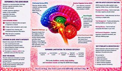
- In ADHD, the brain’s reward system is more strongly driven by what feels interesting, urgent, or immediately engaging, rather than by what is important in the long term.
- This means tasks that are boring, repetitive, or have delayed rewards (e.g., paperwork, admin, long-term projects) can feel much harder to start and sustain, even when the person knows they matter.
- In contrast, tasks that are stimulating, novel, or time-pressured can be completed with high focus and energy.
- As a result, motivation can appear inconsistent—strong in some situations and very low in others—which is often misunderstood as laziness, but actually reflects how the brain processes reward and effort.
Primary functions
- Regulation of motor activity and restlessness
- Action selection and behavioural inhibition
- Habit formation and procedural learning
- Reward processing and reinforcement learning
- Filtering of competing stimuli and responses
Neurobiological basis::
- This system is highly dependent on dopaminergic signalling, particularly within fronto-striatal circuits
- Dopamine modulates the salience of rewards, effort allocation, and the initiation or suppression of behaviour
ADHD-related deficits
- Impaired behavioural inhibition and increased impulsivity
- Reduced capacity to sustain effort for tasks with delayed or abstract reward
- Heightened sensitivity to immediate reward, novelty, urgency, and stimulation
- Difficulty shifting behaviour in response to changing rules or contingencies
- Restlessness or a subjective sense of being internally “driven” to move
Structural and functional findings
- Neuroimaging studies frequently demonstrate reduced volume in basal ganglia structures, particularly the caudate nucleus, in individuals with ADHD
- Functional dysregulation within these circuits contributes to altered reward sensitivity and inefficient action selection

Working Memory
Working Memory
Working memory in ADHD is associated with functional differences across a distributed fronto-striatal–parietal network, including the dorsolateral prefrontal cortex, anterior cingulate cortex, basal ganglia, parietal cortex, and cerebellum. These differences result in impaired maintenance and manipulation of information over time, particularly under conditions of stress, distraction, or cognitive load, and account for core DSM-5 inattentive symptoms observed in adults with ADHD.
Working Memory: Key Brain Areas (and ADHD)
1. Prefrontal Cortex (PFC)
Primary region for working memory
- Especially the dorsolateral prefrontal cortex (DLPFC)
- Responsible for:
- Holding information “online”
- Manipulating information
- Guiding behaviour based on goals and future intentions
ADHD findings
- Reduced activation and efficiency during working memory tasks
- Difficulty sustaining neural firing needed to keep information active
- Highly sensitive to stress, fatigue, and emotional load
Clinical correlate
- Forgetting what one was about to do
- Losing track mid-task
- “Out of sight, out of mind”
2.Anterior Cingulate Cortex (ACC)
Attention control and error monitoring
- Integrates cognition and emotion
- Helps decide:
- What to focus on
- What to ignore
- When effort needs to increase
ADHD findings
- Reduced activation → poor effort regulation
- Difficulty sustaining mental effort over time
- Increased emotional interference with cognition
Clinical correlate
- Inconsistent performance
- Mental fatigue
- Emotional dysregulation worsening forgetfulness
3. Parietal Cortex (especially Posterior Parietal Cortex)
Storage + attentional workspace
- Supports:
- Temporary storage of information
- Shifting and updating attention
- Spatial and sequential working memory
ADHD findings
- Reduced coordination with PFC
- Difficulty updating or refreshing information
- Vulnerability to distraction
Clinical correlate
- Losing track of instructions
- Difficulty juggling multiple steps
- Problems with sequencing and planning
4. Basal Ganglia (especially Striatum)
Gating system for working memory
- Dopamine-dependent “gatekeeper”
- Determines:
- What information gets into working memory
- What gets dropped
ADHD findings
- Dopaminergic dysregulation
- Inefficient gating → either:
- Too much irrelevant information, or
- Failure to hold relevant information
Clinical correlate
- Distractibility
- Mental clutter
- Benefit from stimulant medication (dopamine ↑ → gating improves)
5. Cerebellum
Timing, prediction, and coordination
- Modulates:
- Timing of cognitive processes
- Predictive control
- Automation of routines
ADHD findings
- Structural and functional differences
- Poor temporal coordination of working memory processes
Clinical correlate
- Time blindness
- Poor estimation of task duration
- Difficulty anticipating future steps

Self Management to Time
1. Self-Management to Time
Core Function
Using time to guide behaviour toward future goals.
Russell A. Barkley
’s “Internalisation of Time” Network
- Dorsolateral Prefrontal Cortex (dlPFC)
- Planning
- Holding future goals in mind
- Time estimation
- Fronto-Striatal Circuits
- Coordination of goal-directed behaviour
- Tracking progress toward future rewards
- Cerebellum
- Internal timing
- Temporal sequencing
- Estimation of duration
- Supplementary Motor Area (SMA)
- Timing and sequencing of actions
ADHD Manifestations
- Time blindness
- Chronic lateness
- Poor planning
- Difficulty anticipating future consequences
- Last-minute task completion

Self Organisation
2. Self-Organisation and Problem Solving
Core Function
Planning, organising, sequencing, and solving problems.
Key Brain Regions
Dorsolateral Prefrontal Cortex (dlPFC)
The primary executive control region responsible for:
- Planning
- Organisation
- Working memory
- Complex reasoning
Frontoparietal Network
Includes:
- dlPFC
- Posterior Parietal Cortex
Supports:
- Mental organisation
- Multi-step problem solving
- Attention allocation
Caudate Nucleus
Part of the basal ganglia involved in:
- Cognitive control
- Organising behaviour
- Goal-directed action
ADHD Manifestations
- Disorganisation
- Losing items
- Difficulty managing multiple tasks
- Problems sequencing actions
Self Restraint
3. Self-Restraint
Core Function
Inhibiting impulses and stopping inappropriate responses.
Key Brain Regions
Right Inferior Frontal Gyrus (rIFG)
The brain’s primary inhibitory control centre.
Responsible for:
- Stopping responses
- Suppressing impulses
- Behavioural inhibition
Anterior Cingulate Cortex (ACC)
Monitors:
- Errors
- Conflict detection
- Behavioural adjustment
Basal Ganglia
Particularly:
- Caudate
- Putamen
- Subthalamic nucleus
Supports:
- Response inhibition
- Action selection
ADHD Manifestations
- Interrupting
- Impulsive decisions
- Talking excessively
- Acting without thinking
Self regulation of emotions
5. Self-Regulation of Emotion
Core Function
Managing emotional reactions and recovering from emotional activation.
Key Brain Regions
Amygdala
Responsible for:
- Threat detection
- Emotional salience
- Emotional intensity
In ADHD:
- Often demonstrates increased reactivity
Ventromedial Prefrontal Cortex (vmPFC)
Helps:
- Regulate emotional responses
- Reappraise emotional situations
Anterior Cingulate Cortex (ACC)
Supports:
- Emotional monitoring
- Emotional conflict resolution
Insula
Processes:
- Emotional awareness
- Internal bodily sensations
- Social pain
ADHD Manifestations
- Emotional dysregulation
- Irritability
- Frustration intolerance
- Rejection Sensitivity Dysphoria (RSD)
- Emotional impulsivity
Why Rejection Sensitivity happens
Rejection Sensitivity Dysphoria (RSD) in Attention-Deficit/Hyperactivity Disorder (ADHD)
reflects altered neural processing of :
social threat, emotional salience, and regulation, rather than a discrete diagnostic entity.
It arises from functional and connectivity differences across fronto-limbic, salience, and reward networks.
1. Amygdala: Heightened Threat Detection
The amygdala plays a central role in detecting threat and assigning emotional salience, particularly to social cues such as criticism, exclusion, or perceived disapproval.
- In ADHD, the amygdala demonstrates hyper-reactivity to emotionally salient stimuli
- Neutral or ambiguous interpersonal cues may be misinterpreted as rejection or failure
- This contributes to the rapid onset and intensity of emotional pain characteristic of RSD
Importantly, this response is fast and reflexive, occurring before higher-order cognitive appraisal.
2. Prefrontal Cortex (PFC): Impaired Top-Down Regulation
The prefrontal cortex—particularly the dorsolateral (dlPFC) and ventromedial (vmPFC) regions—modulates emotional responses generated by the limbic system.
In ADHD:
- Reduced PFC activation limits inhibitory control over amygdala output
- Emotional responses are less filtered, less contextualised, and harder to down-regulate
- Individuals may intellectually “know” a response is disproportionate but cannot dampen it in real time
This explains why RSD is often described as overwhelming, uncontrollable, and physically painful.
3. Anterior Cingulate Cortex (ACC): Pain and Social Error Processing
The anterior cingulate cortex integrates emotional pain, cognitive conflict, and social evaluation.
- The ACC is involved in both physical pain and social pain processing
- In ADHD, ACC dysregulation contributes to:
- Intensified distress following perceived rejection
- Heightened sensitivity to interpersonal mistakes or disapproval
- Persistent rumination on social “errors”
This shared circuitry helps explain why rejection in RSD is often described as visceral rather than merely
4. Dopaminergic Reward Pathways: Salience Without Stability
ADHD is associated with dopaminergic dysregulation in fronto-striatal circuits, including the nucleus accumbens.
- Dopamine modulates reward prediction, motivation, and emotional salience
- In ADHD, inconsistent dopamine signalling leads to:
- Over-weighting of negative feedback
- Reduced buffering from prior positive experiences
- Difficulty maintaining emotional equilibrium after criticism
As a result, rejection signals carry disproportionate motivational and emotional weight.
5. Default Mode Network (DMN): Internalisation and Rumination
The default mode network, active during self-referential thought, shows atypical regulation in ADHD.
- Increased DMN intrusion during emotional states promotes:
- Self-blame (“I’ve failed”, “I’m not good enough”)
- Retrospective replay of social interactions
- Prolonged emotional activation after the triggering event
This sustains RSD responses well beyond the initial interpersonal cue.
Integrated Neurobiological Model of RSD in ADHD
RSD reflects the convergence of:
- Hyper-reactive threat detection (amygdala)
- Insufficient top-down emotional inhibition (PFC)
- Amplified social pain signalling (ACC)
- Dopaminergic salience imbalance
- Excessive self-referential processing (DMN)
Together, these systems produce rapid, intense, and enduring emotional responses
to perceived rejection, often without conscious control.
- RSD is not a character flaw or over-sensitivity
- It represents a neurodevelopmentally mediated emotion-regulation vulnerability
- Effective management often requires:
- ADHD-specific pharmacotherapy (to improve fronto-striatal modulation)
- Skills targeting emotional regulation and cognitive reappraisal
- Psychoeducation to reduce shame and self-blame
